No single placement is best for every patient. When there is enough tissue coverage, placing the implant above the muscle allows the breast to move more freely, avoids distortion when the pectoral muscle contracts, and often makes recovery easier. When tissues are thin or the risk of rippling is greater, coverage beneath the muscle can offer an important advantage.
Is it better to place breast implants above or below the muscle?
Above the muscle can be the most natural option when there is enough tissue to cover the implant adequately. The breast moves more freely, does not distort when the pectoral muscle contracts, and surgery is usually less painful. We prefer placement below the muscle when coverage is limited, the edge could be visible or palpable, or the risk of rippling is greater.
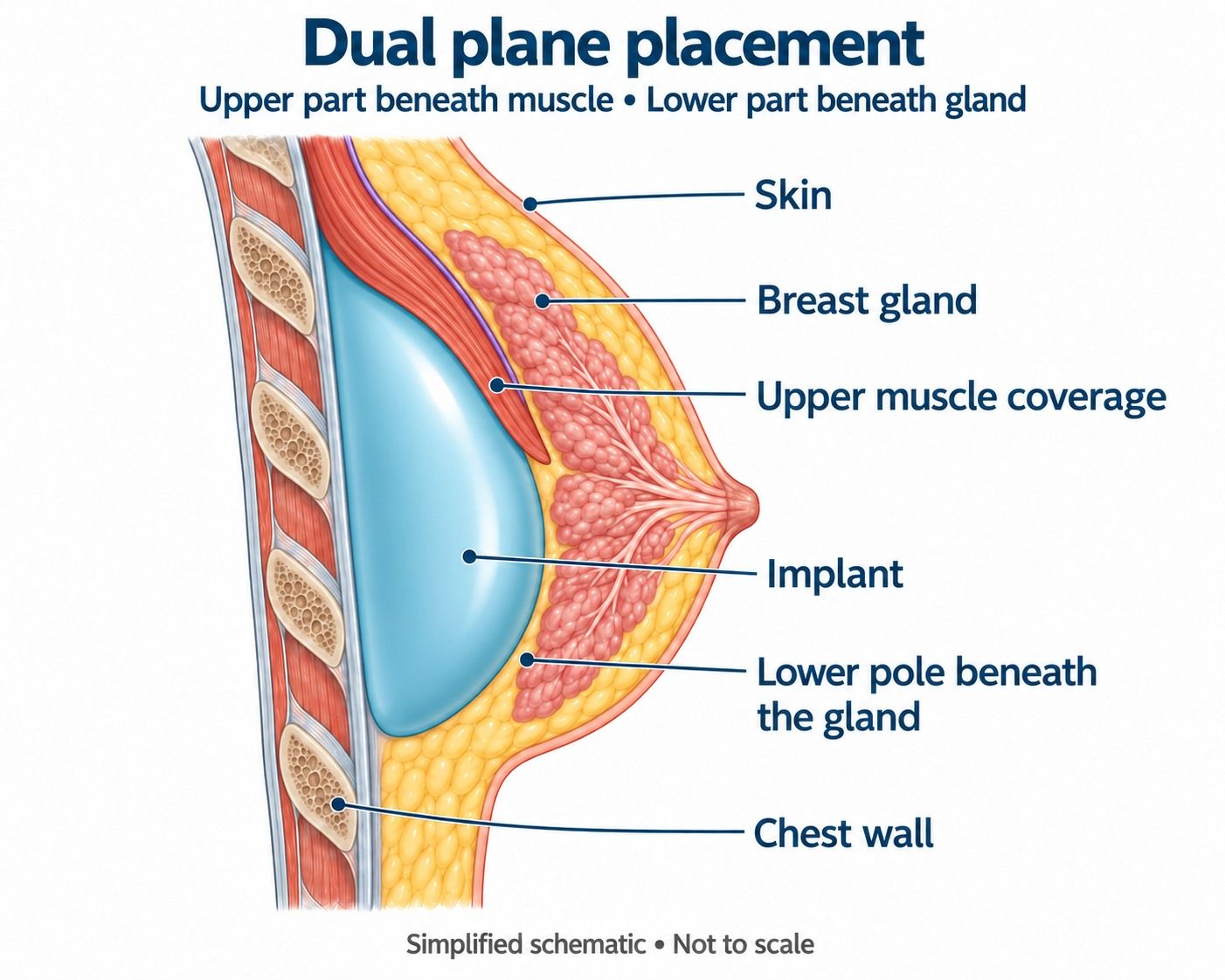
In our practice, the term submuscular includes both complete submuscular placement and dual planetechniques, in which the muscle mainly covers the upper part of the implant. We almost never use complete muscular coverage: we usually perform at least a dual plane I, releasing the lower attachments of the pectoral muscle.
Article contents
Which tissues lie between the skin and the pectoral muscle?
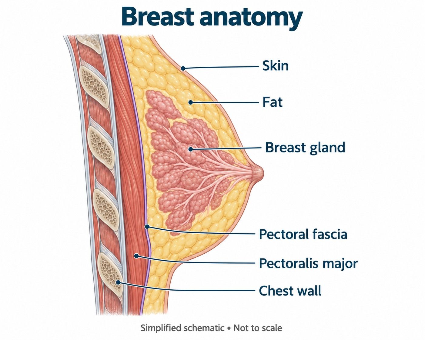
To understand where an implant is placed, it helps to think of the breast in layers. From the surface inward, there are the skin, fat, breast gland, a very thin fascia covering the pectoral muscle, the pectoralis major muscle, and, behind it, the chest wall.
The implant can lie in front of the pectoral muscle—prepectoral placement—or be covered completely or partly by it—submuscular placement. The amount and quality of tissue in front of the implant influence how easily it can be felt, how visible its edge is, and the risk of folds or rippling.
Subglandular, subfascial, and submuscular: the main differences
There are two broad groups. In prepectoralplacements, the implant lies above the muscle; these include subglandular and subfascial placement. In submuscularplacements, the pectoral muscle covers the whole implant or, much more commonly, only part of it using a dual plane technique.
| Placement | Where it sits | Main advantage | Main limitation |
|---|---|---|---|
| Subglandular | Beneath the gland and above the pectoral muscle | Natural movement, no muscular animation, and faster recovery | Needs sufficient coverage to avoid visible edges or rippling |
| Subfascial | Beneath the fascia and above the pectoral muscle | Avoids movement-related distortion, like subglandular placement | The fascia is very thin and, in our experience, adds very little useful coverage |
| Complete submuscular | Implant completely covered by muscle | Maximum muscular coverage | More pain, an initially high implant position, and less natural movement |
| Dual plane | Upper part beneath the pectoral muscle, with the lower part in contact with the gland | Combines upper coverage with better expansion of the lower pole | Can cause animation and takes longer to recover from than prepectoral placement |
What does prepectoral implant placement mean?
Prepectoral means the implant is placed in front of the pectoralis major without using the muscle as coverage. It can lie directly beneath the gland—subglandular—or beneath the pectoral fascia—subfascial.
Both allow the implant to follow the breast's natural movements without being pulled by pectoral contraction. They also avoid muscle dissection, so pain and recovery are usually reduced. A retrospective study of primary augmentation found a shorter duration of pain and faster recovery in the subglandular group than in the subpectoral group, although patients were not randomly allocated. Read the study on PubMed.
≥60 %
Prepectoral placements currently account for at least 60% of our primary breast augmentations.
≈10 %
We use prepectoral placement in approximately 10% of breast lifts with implants.
These are approximate internal figures from our current practice, not trial results or a recommendation that applies to every clinic.
Subglandular implants: above the muscle and beneath the breast
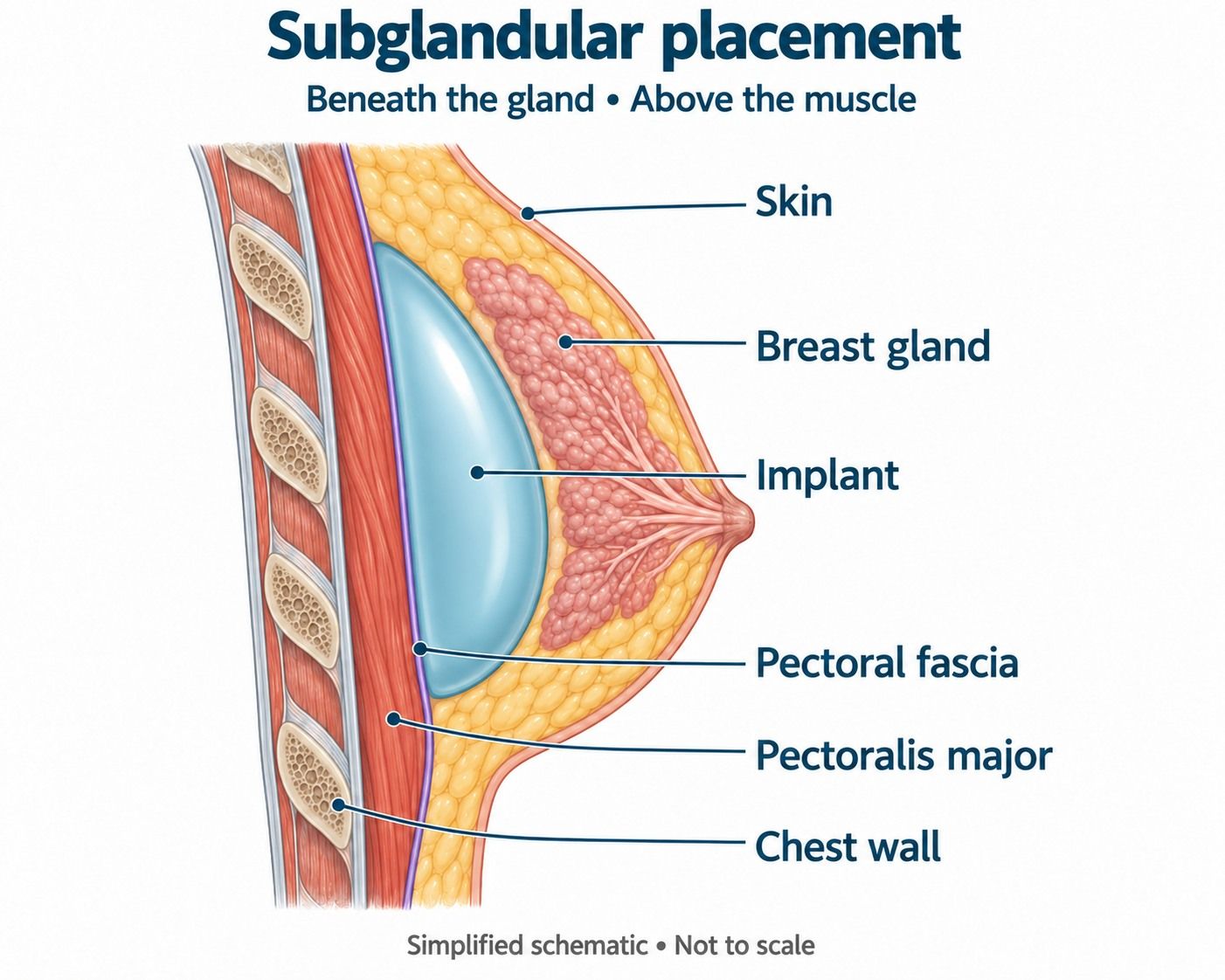
In subglandular placement, the implant lies immediately behind the breast gland and in front of the pectoral muscle. We use it in traditional breast augmentation when coverage is sufficient, and always in our mia® and Preservé™ procedures.
Advantages
- Does not cause breast distortion when the pectoral muscle contracts.
- Allows the implant to follow breast movement more naturally.
- Surgery is usually less painful and recovery is faster.
- Avoids a relatively fixed, high implant becoming separated from a gland that descends over time.
- Can feel very natural with ergonomic implants and adequate coverage.
Limitations
- In very thin patients, the implant edge may be visible or palpable.
- Insufficient coverage increases the risk of visible rippling.
- An implant that is too large or wide can stretch tissues that do not provide sufficient support.
- It is unsuitable if tissue thickness does not allow the implant to be safely concealed.

Subfascial implants: what does the fascia actually add?
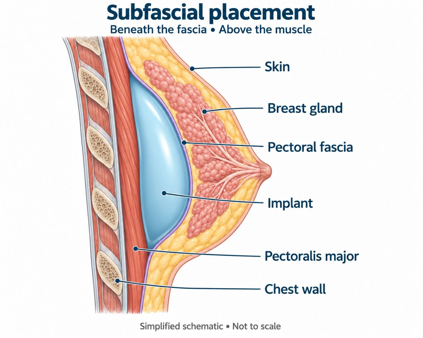
In subfascial placement, the implant sits beneath the fascia covering the pectoral muscle, but remains above the muscle. The fascia is thin: it does not provide partial muscular coverage and should not be described as a thick additional layer.
Some publications have reported less rippling, hematoma, or capsular contracture than with subglandular placement. However, a 2024 meta-analysis noted that all included studies had a high risk of bias. Read the meta-analysis by Yuan and colleagues.
A later analysis found that, when results were separated by implant surface, capsular contracture rates with contemporary smooth implants were similar for subfascial and subglandular placement. This suggests that part of the historical advantage attributed to placement may have reflected the greater use of textured implants in subfascial series. Read Abbott and colleagues.
Submuscular implants: complete and partial coverage
When we describe an implant as submuscular, we include two possibilities: complete muscular coverage, or only the upper part beneath the pectoral muscle using a dual plane technique. The latter is by far the option we use most often.
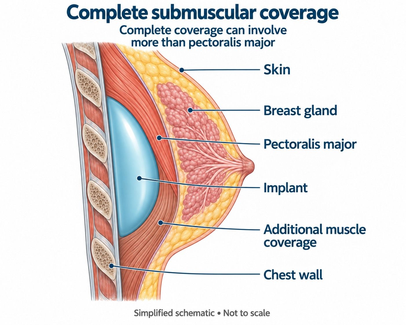
When it can help
- Patients with little fat and glandular tissue in the upper pole.
- Greater risk of a visible edge, palpability, or rippling.
- Certain breast lifts with implants where better implant stabilization is desirable.
- Cases where prepectoral coverage is insufficient for the chosen implant.
The trade-offs
- More pain and slower recovery than with subglandular placement.
- Breast movement or distortion when the pectoral muscle contracts.
- An initial tendency for the implant to sit higher.
- Possible later separation between a high implant and a gland that descends.
Dual plane: a partial submuscular technique
Dual plane is not a category separate from submuscular placement. It is a form of partial submuscular placement combining two maneuvers:
- 1
Release of the lower pectoral attachments. The lower attachments are released so the muscle does not compress the implant or keep it too high.
- 2
Separation of the gland from the muscle. Depending on the type of dual plane, the space between the breast and the pectoral muscle is extended to different degrees.
The aim is to maintain muscular coverage in the upper area—where the edge may be most noticeable—while allowing the lower breast to adapt better to the implant. Tebbetts' original description distinguishes different degrees according to the extent of separation between gland and pectoral muscle. Read the original publication on PubMed.
Because it remains a submuscular technique, dual plane involves working on the pectoral muscle: it generally causes more pain and takes longer to recover from than subglandular placement. Compared with complete muscular coverage, releasing the lower attachments facilitates lower-pole expansion and prevents the muscle from holding the implant excessively high.
Dual plane IRelease of the lower attachments
The lower pectoral border is released without extensive glandular separation. This is the minimum submuscular coverage we usually use.
Dual plane IISeparation up to the lower areola
Greater separation between gland and muscle helps the tissues adapt to the implant.
Dual plane IIIHigher separation
May help breasts with greater laxity or borderline sagging, aiming for the gland and implant to change more harmoniously together.
The exact extent is adapted to the anatomy. These terms describe a surgical strategy; they do not, by themselves, guarantee a particular result.
Beyond the photographA natural breast should also look natural in motion
For many years, surgeons have focused particularly on the static result: front, side, and three-quarter photographs taken while the patient is still. These images are useful, but do not show everything that happens in everyday life.
Women walk, raise their arms, exercise, hug, and contract their pectoral muscles. A submuscular implant may move or distort the contour during contraction. Sometimes the movement is mild; in other cases it produces unnatural animation that does not appear in before-and-after photographs.
A systematic review found that animation deformity is understudied and probably underreported; in the few included studies, its frequency and severity increased with the extent of muscle involvement. Read the systematic review.
Experienced surgeons are also reconsidering subglandular or prepectoral placement when coverage is adequate. This is a change in approach: not a claim that “above” is always better, but a move away from using the muscle out of habit when the patient does not need that coverage. Read the perspective by Montemurro, Gupta, and Mallucci.
How we choose placement for each patient
The choice starts with anatomy, not a favorite technique. We measure coverage, analyze breast and chest shape, assess skin quality, and relate these findings to the implant and the patient's priorities.
Coverage and pinch test
In our protocol, we tend to recommend prepectoral placement when the pinch test and coverage are adequate, usually over 2 cm. This is our own criterion, interpreted alongside the rest of the examination.
Implant and rippling risk
Width, volume, cohesivity, gel behavior, and fit to the chest can affect the risk of visible folds as much as placement.
Pectoral muscle and activity
For patients who train their chest extensively or depend on those muscles, movement-related distortion is particularly important.
Sagging and skin quality
We need to anticipate how the gland, skin envelope, and implant will change—not just how they will look during the first few months.
Primary augmentation or breast lift
We increasingly use prepectoral placements for primary augmentation. In many breast lifts, we prefer muscular coverage to help stabilize the implant.
Informed preferences
The patient should understand the balance between coverage, movement, pain, scars, future surgery risk, and expected appearance.
Mild sagging: better visual centering does not mean lifting the nipple
In a patient with a slightly low nipple or borderline sagging, an implant can develop the lower pole and make the nipple appear better centered relative to breast volume. However, the distance from the sternal notch to the nipple changes little: this is relative centering, or an optical effect, not a true lift.
If the patient wishes to avoid scars for now and understands that further surgery may be needed, we tend to use prepectoral placement or at least a dual plane III. We aim for the implant to follow the gland more closely if tissues descend, avoiding, where possible, a high implant while the breast falls in front of it.
An implant without a breast lift
May be reasonable for borderline patients who accept future sagging and the possibility of needing a lift later. It does not eliminate sagging or guarantee that a second procedure can be avoided.
A breast lift with or without an implant
If the priority is a more stable solution and reducing the likelihood of further surgery in the medium term, we explain that a breast lift addresses skin and nipple position more directly.
3D simulations are particularly useful for these patients. They let us compare what an implant alone can achieve, how much visual centering is realistic, and what would change with a breast lift.
What do we know about contracture, rippling, and other complications?
Studies do not identify one placement that wins on every outcome. A 2026 meta-analysis found higher pooled capsular contracture rates with subglandular than submuscular placement, whereas other problems—such as animation deformity, a high position, or certain malpositions—are more closely associated with muscular coverage. The authors stress individualized selection and note differences between studies. Read the 2026 meta-analysis.
Placement and contracturePooled figures do not predict an individual patient's outcome
Surface, implant generation, technique, contamination, follow-up, and anatomical selection can affect observed rates.
Placement and ripplingCoverage matters, but does not act alone
Muscle can better conceal the upper pole, while gel, size, width, tissues, and dissection influence the rest of the breast.
Placement and movementPectoral contraction changes the result
Prepectoral implants avoid muscular animation; submuscular implants provide coverage at the cost of altered movement.
Placement and recoveryWorking on the muscle has a cost
In our experience and comparative studies, submuscular placement causes more pain and slower recovery than subglandular placement.
Photographs help, but need movement and context
A static image does not show pectoral contraction, feel, mobility, or how the breast behaves when the arms are raised. Nevertheless, comparing cases with similar anatomy, volume, and technique helps explain what each placement can offer.
Frequently asked questions about implants and the pectoral muscle
What does a submuscular implant mean?
It means the pectoral muscle covers all or part of the implant. In our explanation, the term includes both complete submuscular placement and dual plane, or partial submuscular coverage.
Is dual plane the same as below the muscle?
It is a partial submuscular approach. The pectoral muscle mainly covers the upper part, while release of its lower attachments and separation between gland and muscle allow a different relationship with the lower pole.
Which placement is recommended if I have very small breasts?
It depends on actual coverage thickness, not just breast size. When tissue is thin or the pinch test is insufficient, we usually prefer submuscular coverage to reduce palpability and rippling. Examination and simulation help determine the implant and placement.
Do implants below the muscle look more natural?
Not necessarily. They may better conceal the upper edge in thin patients, but can also distort when the pectoral muscle contracts. With adequate coverage, prepectoral placement can provide very natural movement and feel.
Do implants above the muscle cause more rippling?
The risk may increase when coverage is limited, but placement is not the only factor. Implant, width and volume, gel behavior, skin, and precise dissection all matter. With adequate coverage, visible rippling does not necessarily increase.
Are submuscular implants more painful?
Yes. Working on and releasing the pectoral muscle generally causes more pain and slower recovery than subglandular placement. Severity varies with technique, patient, and pain-control protocol.
Can I train my pectoral muscles with submuscular implants?
Many patients can do so after recovery, but contraction may move or visibly distort the implant. For athletes with highly developed pectoral muscles, we give this effect particular attention before choosing placement.
Can dual plane lift a sagging breast?
It does not perform a breast lift. It can develop the lower pole and visually center the nipple in a borderline patient, but does not meaningfully raise its anatomical position. With true sagging, a breast lift is the more direct and stable solution.
Does subfascial placement provide more coverage than subglandular placement?
It adds the fascia, a very thin layer, but not muscular coverage. Some series have reported advantages; in our practical experience, the difference from subglandular placement is small, and some historical results may have been influenced by implant surface.
Medical authorship Specialist in Plastic, Aesthetic and Reconstructive Surgery (Spanish MIR training) Medical registration no. 2828/61062 · Madrid About Breast Implants Above or Below the Muscle: How We Choose: clinical information, safety and expectations. Based on the original Spanish content. Translation does not constitute a new medical review. This information does not replace an individual medical assessment. References support the general medical information. Suitability, alternatives, and risks are discussed individually during consultation.
Medical sources and editorial standards
Selected references for this article
8 references