A seroma is a collection of serous fluid—usually clear and not infected—in a body cavity or between tissues. Surgery is one possible cause, but a seroma can also follow trauma, inflammation, or other conditions that disrupt lymphatic drainage. After breast surgery, ultrasound often shows a thin layer or small collection of fluid around the implant: if it causes no symptoms and is not increasing, it is usually part of normal recovery. However, a breast that enlarges, becomes painful or tense, or develops a clinically significant fluid collection needs investigation and, when appropriate, drainage.
Not every fluid collection seen on ultrasound needs needle aspiration
The decision does not depend simply on whether ultrasound shows fluid. The amount, symptoms, changes over time, wound, presence of an implant, and timing all matter. A small, asymptomatic collection can be monitored; a clinically evident, persistent, or increasing seroma around an implant requires ultrasound, ultrasound-guided aspiration, and sample analysis.
If swelling develops after recovery has ended—months or years later—it is considered a late fluid collection and should be investigated rather than assumed to be benign. Most of these episodes are not malignant, but investigating the cause is the safe way to establish this.
A small collection on ultrasound
Common after surgery, it does not distort the breast or increase, and monitoring is usually sufficient.
Swelling, tension, or persistence
It should be confirmed with ultrasound, aspirated under ultrasound guidance, and sent for microbiological culture.
One breast enlarges months or years later
The fluid, capsule, and implant need investigation, even without fever or severe pain.
In this article
What is a seroma, and why does it develop?
A seroma is a collection of serous fluid—mainly serum and lymph—that builds up under the skin, between surgical tissue planes, or around an implant. Separating tissues during surgery creates an internal space and disrupts small blood vessels and lymphatic vessels. As they heal, inflammation and movement between these surfaces can encourage fluid production.
The size of the space, extent of surgery, individual tissue response, movement, and presence of an implant all influence each case. Even so, there is not always a single identifiable cause.
Serous fluid, usually clear or yellowish. It may be sterile, but a significant collection needs analysis.
A collection of blood, often associated with swelling, tension, pain, and a change in skin color.
May involve warmth, increasing redness, fever, worsening pain, or cloudy, foul-smelling discharge.
These conditions cannot always be distinguished reliably by external appearance or fluid color alone. Examination, ultrasound, and sample collection when indicated help establish the diagnosis.
Early seroma: how much fluid can be normal after surgery?
After breast surgery, ultrasound commonly shows a small amount of fluid around the implant. A thin layer or minimal collection, without distortion, pain, tension, or enlargement, is usually part of the normal tissue response and can be monitored at follow-up appointments.
There is no single volume threshold that determines treatment for every patient. We assess whether the fluid is increasing, the breast is enlarging, the collection is putting pressure on the wound, pain is developing, and an implant is present that could be affected.
Small, stable, and asymptomatic
- Visible only on ultrasound.
- Does not increase breast volume.
- Causes no tension, pain, or wound problems.
- Its progress is documented and monitored.
A clinically significant seroma
- Visible or palpable, and persistent or increasing.
- Causes pain, pressure, distortion, or tension on the wound.
- An implant is present, and the collection may compromise the space around it.
- We perform ultrasound, ultrasound-guided aspiration, and microbiological culture.
Antibiotics do not replace aspiration
For a clinically significant collection around an implant, antibiotics may be indicated if infection is suspected, but they do not remove the fluid or establish its cause. Microbiological testing helps identify or rule out bacterial contamination and guide treatment.
What is a late seroma around a breast implant?
A late seroma is a new fluid collection that develops after the initial recovery period has ended. Many scientific studies use onset at least one year after implant placement as their working definition, but clinically, any new increase in breast volume months or years later warrants assessment.5
It may be associated with inflammation, infection, trauma, implant rupture or wear, capsular changes, or rougher implant surfaces. In some cases, no single cause is identified. A late fluid collection does not, by itself, mean there is a serious disease, but it should not be assumed to be benign without investigation.

If one breast suddenly or progressively enlarges after being stable for some time, arrange a prompt assessment. Do not attempt to puncture or compress it, or start antibiotics or anti-inflammatory medication on your own.
Symptoms of a seroma after breast surgery
Symptoms depend on the amount of fluid, how quickly it accumulates, and the cause. The most common include:
An increase in the size of one breast, often affecting one side or creating asymmetry.
A fluid-like or shifting sensation, pressure, tightness, or a different feeling of weight.
Discomfort around the implant, capsule, or scar.
Changes in breast contour, consistency, position, or firmness.
Clear or yellowish fluid leaking if the incision has not fully closed.
A lump, palpable mass, or enlarged underarm lymph nodes, particularly with late onset.
How is a breast seroma diagnosed and treated?
Ultrasound is the first-choice imaging test to confirm fluid, measure it, examine the capsule, and assess nearby lymph nodes. When an implant is present, it also guides needle aspiration and reduces the risk of damaging the implant.1
Examination and ultrasound
We assess the wound, inflammation, and implant, measure the collection, and look for masses or enlarged lymph nodes.
Ultrasound-guided aspiration
We remove clinically significant fluid and obtain a representative sample without puncturing the implant.
Analysis suited to the situation
A significant early seroma requires microbiological testing. For a late or atypical collection, cytology and immunohistochemistry are added.
Treating the cause
Depending on the results, treatment may include monitoring, repeat aspiration, antibiotics, corticosteroids, a drain, implant replacement, or capsule surgery.

Ultrasound of a fluid collection around an implant
Ultrasound confirms, measures, and documents the collection and, when indicated, guides needle aspiration to analyze or remove the fluid.
Microbiology and follow-up
Culture helps rule out infection around the implant. If the presentation is atypical, the team expands the investigation.
If ultrasound also shows a mass, enlarged lymph nodes, or a skin lesion, investigating the fluid alone is not enough. Spain's medicines and medical devices agency (AEMPS) recommends considering a biopsy and coordinating diagnosis with pathology and the appropriate specialists.
Our approach to fluid around a breast implant
Alongside general recommendations, we use our own protocol based on ultrasound follow-up, accumulated experience in breast surgery, and systematic investigation of late fluid collections. Each decision takes account of the medical history, implant model and age, examination, capsule, and laboratory results.
Routine ultrasound
We use ultrasound during follow-up after breast surgery. It allows us to document small collections before they cause symptoms and check whether they resolve, remain stable, or increase.
Ultrasound-guided aspiration and a specialist laboratory
For a late seroma, we obtain enough fluid for microbiology, cytology, and immunohistochemistry, working with a laboratory experienced in samples from around breast implants.
A tapering course of corticosteroids in selected cases
If there is no infection or evidence of a tumor, the protocol may include a tapering course of corticosteroids to reduce capsular inflammation. This is an individual prescription, never a recommendation to self-medicate.
Implant replacement and capsule treatment
If the seroma returns despite normal test results, we generally consider implant replacement. With implants more than ten years old, even a first benign episode may justify discussing this option.
A tapering course of corticosteroids may be added if a noninfectious inflammatory process is confirmed.
We review the implant and capsule and consider surgery to address the cause.
An implant age over ten years, surface, wear, and patient preferences influence the decision.
Management depends on the diagnosis and is no longer that of a benign seroma.
Implant surface and late seroma
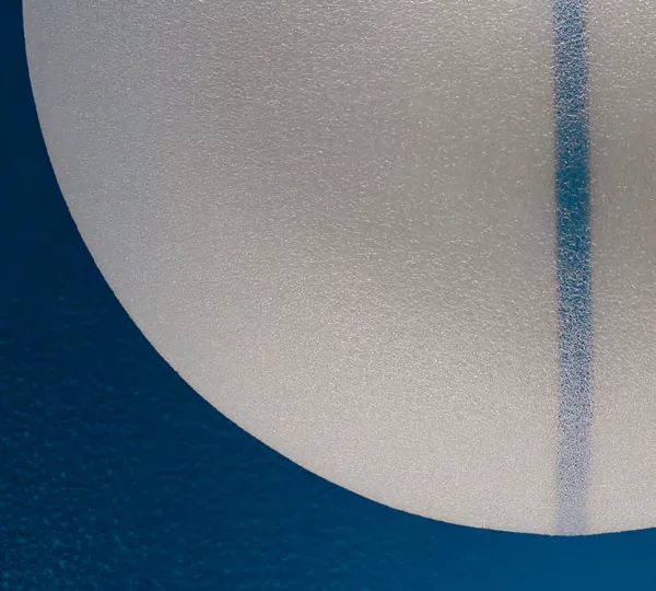
In our experience and in published series, late seromas not caused by lymphoma occur particularly often with rougher implant surfaces. Models implicated include Allergan Biocell and certain Polytech POLYtxt implants in the 20xxx range. This does not mean that every patient with these implants will develop a seroma, or that surface alone can predict the diagnosis.
When replacing implants for this reason, we prefer a much smoother surface—smooth or low-roughness, according to the manufacturer's classification—while individualizing the choice and not presenting any particular brand as a universal solution.
The history of implant surfaces and their indications is discussed in our guide to polyurethane breast implants. This article focuses on the clinical significance of fluid around an implant and does not replace assessment of each patient's specific implant model.
What does en bloc capsulectomy mean?
The capsule is the layer of scar tissue that the body forms around any implant. In Spain, many patients use the term en bloc capsulectomy to mean removing the implant and the entire capsule together as a single piece, without opening the capsule during surgery.
This keeps the fluid contained within the space around the implant and avoids spilling it into the surgical field. When appropriate, the complete specimen is sent for pathological examination of the capsule, fluid, and any suspicious areas.
One piece, without opening the compartment. Whether this can be achieved depends on the anatomy, how firmly the capsule is attached, and the safety of neighboring tissues.
International terminology is not uniform. The 2024 BSCC consensus calls complete removal as one piece without a margin total intact capsulectomy and reserves en bloc for removal with a margin of healthy tissue when an implant-associated cancer is suspected or confirmed.7 We explain both terms to avoid misunderstandings, while also describing the common Spanish use of "en bloc capsulectomy."
Not every implant removal requires a complete or en bloc capsulectomy. The indication and risks depend on the diagnosis, capsule, implant plane, and proximity to the ribs, muscle, and pleura. When operating for recurrent seroma or a capsular abnormality, we aim to remove the capsule completely whenever it is technically safe.
Late seroma, BIA-ALCL, and other capsular tumors
Breast implant-associated anaplastic large cell lymphoma (BIA-ALCL, known as LACG-AIM in Spanish) is a rare disease of the lymphatic system that develops in the fluid or capsule surrounding the implant. It is not cancer of the breast gland. It has mainly been associated with textured implants, and its most common presentation is persistent swelling of one breast that develops long after surgery.1
Most late seromas are not lymphoma. Nevertheless, AEMPS recommends ultrasound and, if fluid is present, aspiration and submission for microbiology and pathology/cytology, informing the laboratory of the suspected diagnosis. Testing includes cell morphology and, when appropriate, CD30 and ALK markers.1
A lymphoma of the capsule
It may present with a late fluid collection, a mass, hardening, pain, skin changes, or enlarged lymph nodes. If confirmed, the patient is referred to a multidisciplinary team.
Even more exceptional
The FDA has reported squamous cell carcinoma and other capsular tumors. These are extremely uncommon, and a reliable incidence and all their risk factors are not yet known.4
This information does not justify removing implants in patients without symptoms
The FDA does not recommend implant removal solely because of concern about these tumors in a patient without symptoms. The reasonable approach is to maintain recommended follow-up and investigate any new change. If a capsular disease is confirmed, treatment should be planned with a multidisciplinary team.4
Can a seroma be prevented after surgery?
Not all seromas can be prevented. Planning, careful technique, control of bleeding, minimizing internal spaces, and selective use of drains are measures the surgeon adapts to the procedure and individual risk.
After surgery, it is important to follow instructions about your bra, activity, wound care, and appointments. Avoiding strenuous activity that has not been approved during recovery may reduce movement and tension on the tissues, but a seroma should not automatically be blamed on something the patient did wrong.
Drains have specific indications and do not guarantee that fluid will not develop after they are removed. Our guide to drains in breast surgery explains when we use them and why they are not routine in every augmentation.
Have you noticed a recent change, or do older implants need assessment?
My breast has enlarged or is painful
Arrange a prompt assessment
Especially if the change affects one side, is progressing, involves fever, or includes fluid leaking from the wound.
Contact the clinic
My implants are older, or the seroma has returned
Assess the implant, capsule, and possible replacement
We review tests, model, surface, implant age, and surgical alternatives individually.
Request an implant assessment
If you develop fever, feel unwell, or have severe pain or rapid swelling outside clinic hours, seek care at an emergency department. A website form must not delay urgent medical care.
Questions about fluid and seroma after breast surgery
Is fluid around an implant normal after surgery?
Can a seroma resolve on its own?
How is a seroma aspirated without damaging the implant?
Does clear or yellow fluid mean there is no infection?
Can antibiotics cure a seroma?
When is a seroma considered late?
Is every late seroma a lymphoma?
What tests are performed on fluid from a late seroma?
What happens if the findings are benign?
What happens if the seroma returns?
Are en bloc and total capsulectomy the same?
Should implants be removed in patients without symptoms because of concern about BIA-ALCL or BIA-SCC?
Can a seroma contribute to capsular contracture?
When should I seek emergency care?
Protocols, consensus statements, and studies used
- AEMPS: clinical protocol for detecting breast implant-associated ALCL, version 2.
- FDA: questions and answers about BIA-ALCL.
- NCBI Bookshelf: postoperative seroma management.
- FDA: update on squamous cell carcinoma and other capsular tumors.
- Bengtson et al.: consensus on late fluid collections around breast implants.
- Marcelli, Aso et al.: comparison of POLYtxt and MESMO focusing on late seroma.
- BSCC: consensus on capsulectomy terminology and management.
- AEMPS: information and monitoring of breast implant-associated ALCL.
A small collection on ultrasound may be normal; a clinically evident or late seroma needs investigation
There is no need to be alarmed by every tiny amount of fluid, or to accept an enlarging breast as normal. Ultrasound distinguishes an expected finding from a significant collection and allows precise aspiration.
For a late seroma, analyzing the sample is as important as removing the fluid. Only after the results are known can the team decide between monitoring, anti-inflammatory treatment, antibiotics, implant replacement, or capsule surgery.
Medical authorship Specialist in Plastic, Aesthetic and Reconstructive Surgery (Spanish MIR training) Medical registration no. 2828/61062 · Madrid About Seroma After Breast Surgery: Causes, Symptoms, and Treatment: clinical information, safety and expectations. Based on the original Spanish content. Translation does not constitute a new medical review. This information does not replace an individual medical assessment. References support the general medical information. Suitability, alternatives, and risks are discussed individually during consultation.
Medical sources and editorial standards
Selected references for this article
8 references