Yes: you can have mammograms with breast implants and should follow the screening program appropriate for your age, country and individual risk. Tell the imaging center when booking so an experienced team can take additional views that move the implant back and show as much breast tissue as possible.
Implants do not prevent screening, but they change how the mammogram is performed
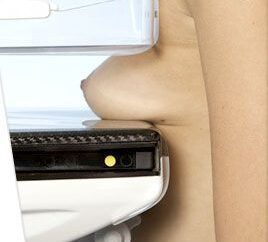
The implant is visible on the mammogram and can obscure some tissue behind it. In addition to standard images, the team usually takes implant-displacement views, also called Eklund views. The implant is gently moved toward the chest wall while the breast tissue is brought forward for imaging.1
Ultrasound and MRI do not automatically replace mammography. They answer complementary or different questions. The choice depends on whether you need routine cancer screening, investigation of a new symptom or assessment of silicone-implant integrity.
Screen for cancer without symptoms
Mammography or breast tomosynthesis remains the basis of screening according to age and risk.
Investigate a symptom
A lump, discharge, retraction or change in breast size calls for a targeted diagnostic assessment.
Check the implant
Ultrasound and non-contrast MRI are better suited to assessing possible silicone-implant rupture.
In this article
How is a mammogram performed when you have implants?
Tell the center about your implants when booking and remind the imaging team before the examination. This allows time for the adapted protocol. If you know your implant type and its position relative to the chest muscle, share that information and bring previous images if available. Choosing a center experienced with augmented breasts is sensible.2
Standard views
The usual mammographic images assess both breasts and the implants’ position.
Implant-displacement views
Four additional images—two per breast—are commonly taken with the implant moved back and the breast tissue drawn forward.
Specialist interpretation
The radiologist considers these images alongside breast density, previous studies, examination findings and any symptoms.
Can the entire breast always be seen?
No. An implant can obscure some tissue, and dense breast tissue can also reduce mammographic sensitivity. Displacement views improve visualization, but no screening test has 100% sensitivity. The amount of tissue, capsular contracture and implant position may affect how easily the views can be obtained.
Mammogram, ultrasound or MRI: what is each used for?
| Test | Common use | Important distinction |
|---|---|---|
| Mammography or tomosynthesis | Breast-cancer screening and diagnostic assessment of selected findings. | Additional views are needed with implants. Mammography may show some ruptures, but a normal result does not rule out silicone-implant rupture. |
| Breast ultrasound | Assessing a lump, fluid or lymph nodes; complementing mammography; and initially checking implant integrity. | It uses no ionizing radiation, but does not generally replace routine mammographic cancer screening. |
| MRI without contrast | Assessing silicone-implant integrity, especially if there are symptoms or ultrasound is inconclusive. | This is the most sensitive method for a silent rupture; the implant protocol does not require gadolinium contrast.4 |
| MRI with contrast | Selected cancer-related questions, including assessment of people at high risk. | It answers a different question from MRI performed solely to check an implant for rupture. |
A normal screening mammogram does not end the investigation of a new lump. The breast team decides whether diagnostic mammography, ultrasound, MRI or biopsy is needed.
What imaging do we request before breast surgery?
The aim is to assess the breast appropriately for the patient’s age, tissue, history, symptoms and planned operation. There is no single test for everyone.
Mammogram and/or ultrasound
Our clinic may request mammography, ultrasound or both according to breast tissue, personal and family history, symptoms and recent imaging.
Individual assessment
We consider history, examination, the planned procedure and whether recent imaging is still suitable.
When a mammogram is indicated, we aim to obtain it shortly before surgery. This provides a recent baseline and, if there are no symptoms or other medical indications, may allow routine imaging to be deferred while the breast settles after surgery.
Mammography and/or ultrasound according to individual assessment.
We generally aim to defer a routine mammogram until then when there is no reason to perform it sooner.
Diagnostic needs take priority over any routine postoperative timetable.
Screening ages vary by country and individual risk. Spain approved a gradual expansion of its population program to ages 45–74 in 2026; this is not a rule for patients living in the UK or elsewhere in Europe. Follow the screening invitations and risk-based advice in your home healthcare system.3
How often should implants be checked with ultrasound or MRI?
Checks for possible implant rupture do not replace breast-cancer screening. These schedules may overlap, but they serve different purposes.
Annual clinical review
Spanish patient information updated in July 2026 recommends annual review with a plastic surgeon while implants remain in place, ultrasound for implant carriers and MRI when needed.4
Ultrasound or MRI from years 5–6
For asymptomatic silicone implants, guidance recommends a first ultrasound or non-contrast MRI at 5–6 years, then one of these tests every 2–3 years. Symptoms or an uncertain ultrasound may warrant MRI sooner.56
Annual ultrasound, with MRI around ten years
We generally perform or request an annual ultrasound. If the examinations remain reassuring, we may suggest MRI around ten years for a more detailed implant assessment. We bring it forward for symptoms, significant trauma, inconclusive ultrasound or suspected rupture.
This approach is compatible with US guidance, which accepts ultrasound or MRI as alternatives for periodic implant checks rather than requiring both each time. Our annual ultrasound is more frequent; MRI adds detail when clinically indicated.
Can a mammogram rupture an implant?
The risk is very low, but it is not zero. Regulators and imaging services recognize the possibility of damage from compression, although reporting systems cannot provide a precise rate. This is not a reason to abandon appropriate cancer screening. Tell the team about your implants and choose a center experienced in imaging patients with them.411
I have seen a small number of cases compatible with damage during mammography, generally involving old or already deteriorated implants. That experience cannot establish a risk rate or prove the implant was intact before compression.
Significant capsular contracture can make displacement views harder and more uncomfortable. Report marked hardness, a change in shape or suspected rupture before imaging so the radiologist can choose the most appropriate assessment.
Do implants delay cancer detection or worsen prognosis?
Silicone implants have not been shown to increase the risk of ordinary breast-gland cancer.4 They can obscure some breast tissue on a mammogram and reduce sensitivity, especially if additional views are not taken.
Lower mammographic sensitivity
A large cohort found lower screening sensitivity among women with augmentation, but did not find worse prognostic characteristics in the cancers detected.7
Series of 4,082 patients
Women with implants had more palpable lesions and more false-negative mammograms, without significant differences in stage, tumor size, recurrence or survival in that series.8
Observational evidence has limitations
A 2013 review raised a possible survival concern but noted inadequate adjustment for confounding factors. A 2026 review did not establish a worse prognosis, while emphasizing the need for optimized imaging.910
The useful conclusion is not that implants make no difference
The more responsible conclusion is: implants do not increase the usual breast-cancer risk or remove the need for screening, but imaging must be adapted and every new symptom assessed. Continuing screening, sharing prior studies and using experienced centers help prevent avoidable delays.
Some studies suggest a lump may be easier to feel when breast tissue lies over an implant. This is not a protective effect and never replaces imaging or medical assessment.
Breast symptoms with implants: what should be assessed?
Warning signs are largely the same with or without implants. An implant adds other possible explanations—contracture, rupture, fluid or displacement—but a new change should not be dismissed as “just the implant” without assessment.
In the breast, near the implant capsule or in the armpit, even after a recent normal mammogram.
New retraction, bloody discharge, a persistent skin change or a wound that does not heal.
Late swelling on one side, enlargement or fluid around an implant.
Progressive hardening, a new deformity, pain or a clear change since previous checks.
These changes do not necessarily mean cancer, but they need examination and targeted diagnostic imaging. Sudden late swelling, fluid or a mass near the capsule also requires assessment for implant-associated disease. Implant-associated lymphoma arises in the capsule and is not ordinary breast-gland cancer.
What if implants were placed after a mastectomy?
Follow-up after breast reconstruction cannot simply copy the schedule used after cosmetic augmentation. It depends on whether one or both breasts were removed, what tissue remains, the type of reconstruction, cancer history and the treating breast team’s plan.
After bilateral mastectomy, routine mammography of reconstructed breasts may not be indicated. If a natural breast remains after unilateral mastectomy, it usually continues the appropriate screening program. The treating breast team should set the individual imaging plan.
Are you considering surgery, or do you already have implants?
I am considering augmentation
Plan surgery and preoperative imaging
We assess your anatomy, history, simulation and any breast imaging needed.
Request an augmentation consultation
I already have implants
Check the implant and surrounding tissue
We review ultrasound findings, symptoms, changes in shape and whether further assessment is needed.
Request an implant-revision consultation
If you have a lump, nipple discharge, skin retraction or late swelling on one side, seek diagnostic assessment from a primary-care clinician, radiologist, gynecologist or breast unit first. An aesthetic-surgery enquiry must not delay that assessment.
Mammograms, breast cancer and implants
Can I have a mammogram with silicone implants?
Could a mammogram rupture an implant?
Does ultrasound replace mammography?
Does MRI replace mammography?
Can a mammogram show a ruptured implant?
Do implants increase breast-cancer risk?
Can cancer be hidden behind an implant?
What if I feel a lump and have implants?
What imaging is requested before augmentation?
When can I have a mammogram after surgery?
How often do you check implant integrity?
Is follow-up the same after cancer reconstruction?
Guidelines and studies consulted
- American Cancer Society: mammograms for women with breast implants.
- FDA: frequently asked questions about mammograms and implants.
- Spanish Ministry of Health: expansion of breast-cancer screening to ages 45–74.
- AEMPS: patient information on breast implants, updated July 2026.
- FDA: ultrasound and MRI follow-up recommendations.
- American College of Radiology: breast implant evaluation, updated 2025.
- Miglioretti et al.: mammographic accuracy and cancer characteristics.
- Handel: effects of implants on diagnosis, prognosis and treatment.
- Lavigne et al.: observational systematic review and meta-analysis.
- 2026 review of implants, cancer detection and oncological outcomes.
- FDA: reports of adverse events during mammography.
Having implants does not mean choosing between them and early cancer detection
Mammography remains possible and important when indicated. Tell the imaging team, use an adapted technique and distinguish tests for breast tissue from tests for implant integrity.
If you are considering surgery, we plan appropriate preoperative imaging. If you already have implants, we can discuss checks of their integrity. If a new breast symptom appears, diagnostic assessment takes priority over the next routine review.
Medical authorship Specialist in Plastic, Aesthetic and Reconstructive Surgery (Spanish MIR training) Medical registration no. 2828/61062 · Madrid About Can You Have a Mammogram With Breast Implants? Screening and Safety: clinical information, safety and expectations. Based on the original Spanish content. Translation does not constitute a new medical review. This information does not replace an individual medical assessment. References support the general medical information. Suitability, alternatives, and risks are discussed individually during consultation.
Medical sources and editorial standards
Selected references for this article
11 references